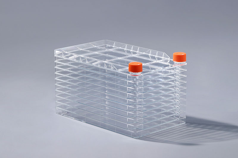
Four detection methods for mycoplasma contamination in cell factories
Mycoplasma contamination is a very common problem when culturing cells in cell factories. Unlike other contaminations, mycoplasma-contaminated cells generally do not become cloudy, so it is difficult to judge with the naked eye. There are four ways to determine whether cells are contaminated with mycoplasma:
1. DNA fluorescence staining
The DNA fluorescence staining method is based on the principle that the fluorescent dye Hoechst33258 can bind to the AT base-rich region in the DNA of mycoplasma. Fluorescent dots are mycoplasma DNA rich in AT base regions.
2. PCR technology
The specific primers are designed according to the conserved sequences in the Mycoplasma genome, the nucleic acid of the sample to be tested is amplified, and the diagnosis is made by analyzing the size of the amplified product. PCR detection technology is used for the detection of mycoplasma contamination, with short cycle, high sensitivity, good specificity, simple operation, and can detect a large number of samples at one time.
3. Enzyme-linked immunosorbent assay (ELISA)
ELISA is used in the detection of mycoplasma contamination, with good specificity and sensitivity, and can complete the detection of a large number of samples at one time. It has the characteristics of simple, rapid and quantitative qualitative detection.
4. Electron microscope
Generally, cells in cell factories are cultured for 48 to 72 hours. Before the cells are nearly confluent, the cells are digested with trypsin to make a cell suspension, fixed, embedded, and sliced before observation.
The above are four commonly used detection methods when detecting mycoplasma contamination in cell factories. At present, there are more than 20 kinds of mycoplasma known to contaminate cells, of which more than 95% are oral mycoplasma, so operators should pay attention to aseptic operation when culturing cells.

评论
发表评论